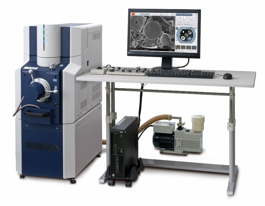
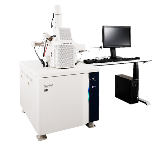
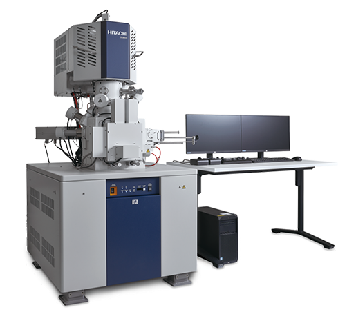
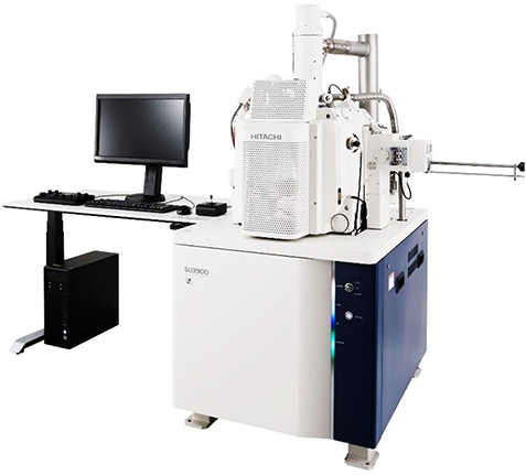
SEM SU3900
Product Description
Hitachi High-Tech's scanning electron microscopes SU3800/SU3900 deliver both operability and expandability. The operator can automate many operations and efficiently utilize their high performance. The SU3900 is equipped with a large multipurpose specimen chamber to accommodate observation of large samples.
Specification
| General Characteristic | |
|---|---|
| Secondary Electron Resolution | 3.0 nm (accelerating voltage 30 kV, WD=5 mm, high vacuum mode) 15.0 nm (accelerating voltage 1 kV, WD=5 mm, high vacuum mode) |
| Backscattered Electron Resolution | 4.0 nm (accelerating voltage 30 kV, WD=5 mm, low vacuum mode) |
| Magnification | ×5 to ×300,000 (magnification of image*1) ×7 to ×800,000 (magnification of actual display*2) |
| Accelerating Voltage range | 0.3 kV to 30 kV |
| Low Vacuum Mode Setting | 6 to 650 Pa |
| Image Shift | ± 75 µm (WD=10 mm) |
| Max. specimen size | Φ 300 mm |
| Specimen stage | X : 0 to 150 mm Y : 0 to 150 mm Z : 5 to 85 mm R : 360° in continuous mode T : -20 to +90° Max. Movable Range : Φ 200 mm (in combination with R) Max. Movable Height : 130 mm (WD= 10 mm) Motor Drive : 5-axis motor drive |
| Electron Optics | Electron-Gun : Pre-centered cartridge type tungsten hairpin filament Objective-Lens Aperture : 4-hole movable aperture Detectors : Secondary electron detector, sensitive semiconductor backscattered electron detector WD for EDX analysis : WD = 10mm (T.O.A = 35°) |
| Image display | Auto-Axis Alignment Function Beam control : auto (AFS→ABA→AFC→ABCC) Optical axis adjustment: auto (current alignment) Beam brightness: auto Auto Image Adjustment Function Auto brightness and contrast control (ABCC) Auto focus control (AFC) Auto stigma and focus (ASF) Auto filament saturation (AFS) Auto beam alignment (ABA) Auto start (HV-ON→ABCC→AFC) |
Resources
| Scanning Electron Microscope SU3800 & SU3900_1.pdf |
|---|